
PRODUCTS
iClear™(Medical Image Enhancing Solution)
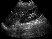
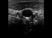
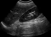
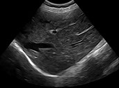
iClear™is an image enhancement solution for medical imaging systems such as ultrasound, X-ray, CT, MRI, and mammography. It reduces noise and enhances edges in real time, improving contrast, resolution, and overall image clarity.
We provide multiple filter sets with different enhancement levels, optimized for the characteristics of our partners' medical imaging systems.


Original Image
Enhanced Image
Original Image
Enhanced Image
Original Image
Enhanced Image
<
>

Original Image
Original Image
Enhanced Image
Enhanced Image
Original Image
Enhanced Image
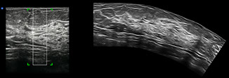
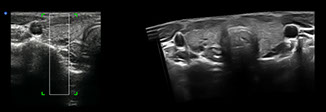
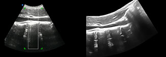
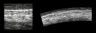
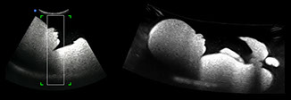
iPano™(Ultrasound Panoramic Imaging Solution)
iPano™ is an ultrasound imaging technique that stitches multiple B-mode images together to create a single composite image with an increased field of view. It provides visualization of anatomical structures and their relationships in a single large field-of-view image, making it highly useful for diagnosis in musculoskeletal imaging, orthopedics & rehabilitation, obstetrics & fetal examination, vascular & neurology, and more.
Result panorama images on the right are after speckle reduction and edge enhancement by iClear™.
Breast
Liver
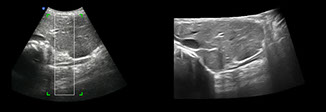
Liver and IVC
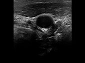
Thyroid-multinodular thyroid
Breast
Thyroid-multinodular thyroid
Liver
Liver and IVC
Leg-long axis scan
36weeks fatal phantom
<
>
Leg-long axis scan
36weeks fatal phantom
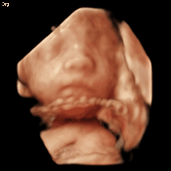
iClear3D™
iClear3D™ AI engine analyzes the contours and surfaces of structures visible in 3D images to accurately identify them. It then enhances the intensity and continuity of weaker signals that form these contours and surfaces, making them more distinct. Meanwhile, irregular surfaces and contours generated by noise in 2D images are smoothed and refined by weakening inconsistent signals, ultimately improving the overall quality of the 3D image.
By performing these high-precision enhancements automatically, without requiring additional user input, iClear3D™ AI delivers clearer imaging results in daily ultrasound scanning without extra manual adjustments. This efficiency optimizes workflow for medical professionals while enhancing overall satisfaction for both doctors and patients.
4D Ultrasound Volume Imaging Solution
Surface Rendering : The surface of the target object, shown in grayscale in the 2D image, is rendered into 3D, while liquid materials displayed in black in the 2D image are rendered as transparent, making them invisible in the 3D image
Depthview Rendering : To enhance the distinction between near and far structures within 3D volume data on the monitor, the system utilizes a dual-color approach. The near structure is displayed in a user-selected color from the 3D palette, while the far structure is represented in blue or dark grey, as illustrated below.

Surface Rendering Depth Rendering

Surface Rendering
Depth Rendering
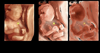
Realskin Rendering : To achieve a realistic skin effect, the system considers the location of the light source relative to the 3D volume and calculates its impact on the fetal surface structure. Users have the flexibility to freely adjust and reposition the light source around the 3D volume data.

Realskin Rendering with Left Realskin Rendering with
Light Source Positioning. Front Light Source Positioning

Realskin Rendering with Left Light Source Positioning.
Realskin Rendering with
Front Light Source Positioning.
Silhouette Rendering : Users can examine both the internal and external structures of the target object in the 3D volume by adjusting the transparency level. Increased transparency reveals more internal structural details with reduced surface visibility, while decreased transparency emphasizes the surface with fewer internal details visible.

Silhouette Rendering with Silhouette Rendering with Silhouette Rendering with
Transparency Level 0 Transparency Level 50 Transparency Level 50
(Feont Light Source Positioning) (Front Light Source Positioning) (Back Light Source Positioning)
Silhouette Rendering with
Transparency Level 0
(Front Light Source Positioning)
Silhouette Rendering with
Transparency level 50
(Front Light Source Positioning)
Silhouette Rendering with
Transparency Level 50
(Back Light Source Positioning)
GALLERY
4D Ultrasound Volume Imaging Solution


iClaer3D™


Tel. +82-53-652974 | Fax. +82-53-652-9765
Copyright 2025 ONDEMANDSOFT ALL rights reserved.